
Imaging the dynamics of fertilization, from mouse to human
Fertilization is a highly dynamic process, yet our knowledge about this crucial event derives mostly from analyses of static samples. We recently advanced the use of live-imaging to capture the events immediately after sperm entry in mouse and human oocytes.
Scheme of fertilization and preimplantation development

Capturing the rapid dynamics of sperm entry into a mouse oocyte. The zona pellucida is intact, but not visualized here. One spermatozoon (blue) enters the sperm from the top region. The oocyte then completes meiosis (see polar body extrusion in the bottom region)

The sperm pauses under the oocyte cortex and undergoes extensive decondensation of its DNA

Time-frames show sperm decondensation at high resolution

The fertilization cone (FC), a protrusion above the sperm DNA, forms far from the initial sperm site (SEP)


Imaging the actin cortex (orange) uncovers massive actin flows triggered by sperm entry
Actin flows pull cortex-associated structures including the sperm DNA towards the cytokinetic furrow
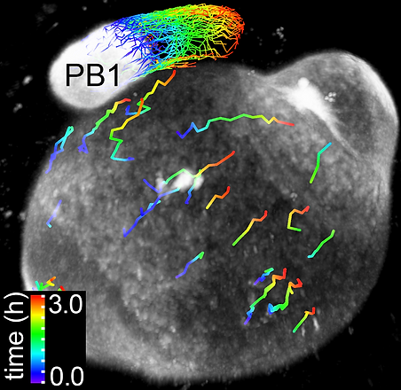
Capturing the dynamics of sperm entry into a human oocyte

Detailed analyses of sperm entry dynamics and movement inside the mouse oocyte

Fixed sample showing sperm on left side and maternal chromosomes completing meiosis on the right side
